38 human cell with labels
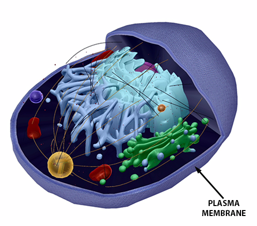
Human Cell Diagram, Parts, Pictures, Structure and Functions Diagram of the human cell illustrating the different parts of the cell. Cell Membrane. The cell membrane is the outer coating of the cell and contains the cytoplasm, substances within it and the organelle. It is a double-layered membrane composed of proteins and lipids. The lipid molecules on the outer and inner part (lipid bilayer) allow it to ... human body | Organs, Systems, Structure, Diagram, & Facts The cell is the basic living unit of the human body—indeed, of all organisms. The human body consists of trillions of cells, each capable of growth, metabolism, response to stimuli, and, with some exceptions, reproduction. Although there are some 200 different types of cells in the body, these can be grouped into four basic classes.
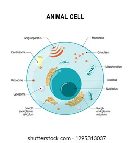
A Labeled Diagram of the Animal Cell and its Organelles A Labeled Diagram of the Animal Cell and its Organelles There are two types of cells - Prokaryotic and Eucaryotic. Eukaryotic cells are larger, more complex, and have evolved more recently than prokaryotes. Where, prokaryotes are just bacteria and archaea, eukaryotes are literally everything else.

Human cell with labels
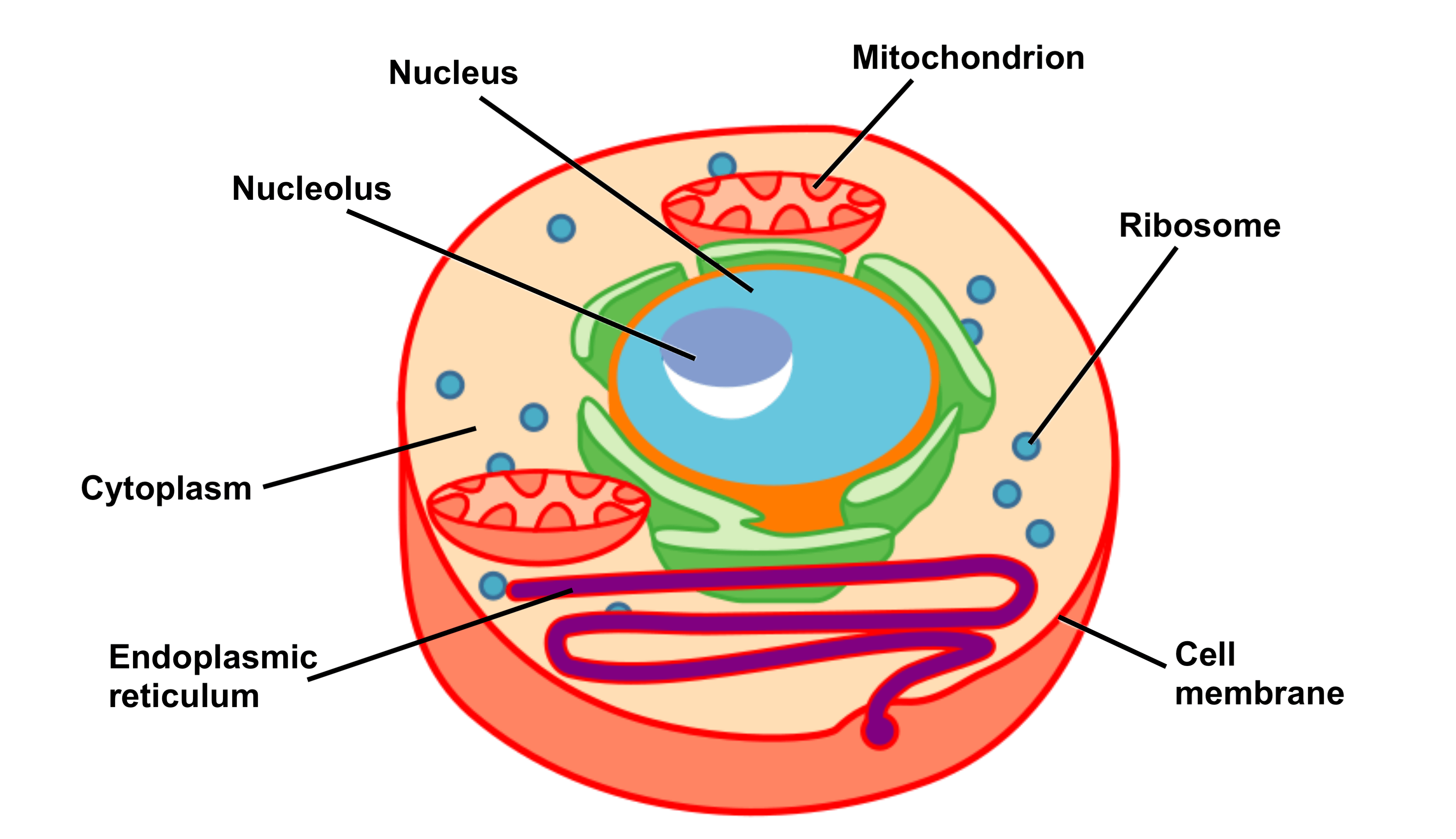
Label Human Cell Teaching Resources | Teachers Pay Teachers Human Cells and Blood Cells ; Label the Various Cells Diagram:This is a great supplement for students to review/assess and strengthen their knowledge the on the TYPES HUMAN CELLS UNIT. Answer key is included. Colorful and Black and White versions of worksheet is included.It includes total ONE worksheet. generalized human cell, labeled Diagram | Quizlet Start studying generalized human cell, labeled. Learn vocabulary, terms, and more with flashcards, games, and other study tools. Scheduled maintenance: Saturday, September 10 from 11PM to 12AM PDT Human Physiology - Cell structure and function Cell, or Plasma, membrane - encloses every human cell Structure - 2 primary building blocks include protein (about 60% of the membrane) and lipid, or fat (about 40% of the membrane). The primary lipid is called phospholipid , and molecules of phospholipid form a 'phospholipid bilayer' (two layers of phospholipid molecules).
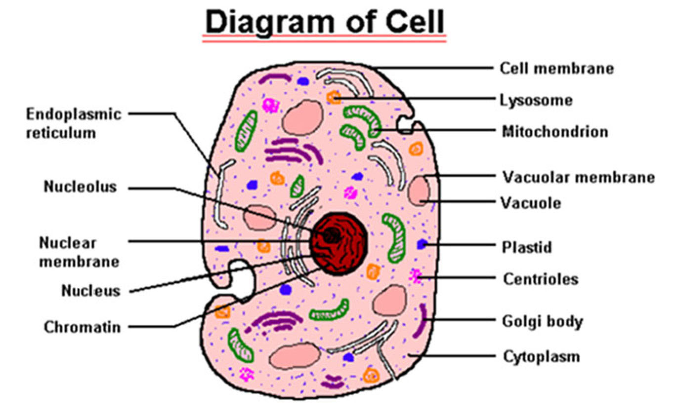
Human cell with labels. Free Printable Plant and Animal Cells Worksheets - Homeschool Giveaways Label the Parts of an Animal Cell Printable Label and Color the Parts Animal Cell - This animal cell handout is perfect for students studying the different parts of the cell including the SER (Smooth Endoplasmic Reticulum), Golgi Bodies, and more. Human Cell Diagram Pictures, Images and Stock Photos Diagram of generic plant and animal cells, showing major organelles including nucleus, nucleolus, rough endoplasmic reticulum, smooth endoplasmic reticulum, cell membranes, golgi apparatus, mitochondria, vacuoles, lysosomes, ribosomes, and centrioles. The plant cell obviously also has a cell wall and chloroplasts. PDF Human Cell Diagram, Parts, Pictures, Structure and Functions Human Cell Diagram, Parts, Pictures, Structure and Functions The cell is the basic functional in a human meaning that it is a self-contained and fully operational living entity. Humans are multicellular organisms with various different types of cells that work together to sustain life. Other non-cellular components in the body include water ... What is a cell?: MedlinePlus Genetics What is a cell? Cells are the basic building blocks of all living things. The human body is composed of trillions of cells. They provide structure for the body, take in nutrients from food, convert those nutrients into energy, and carry out specialized functions. Cells also contain the body's hereditary material and can make copies of themselves.
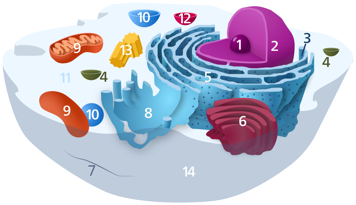
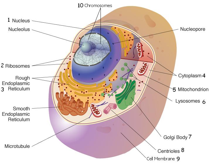
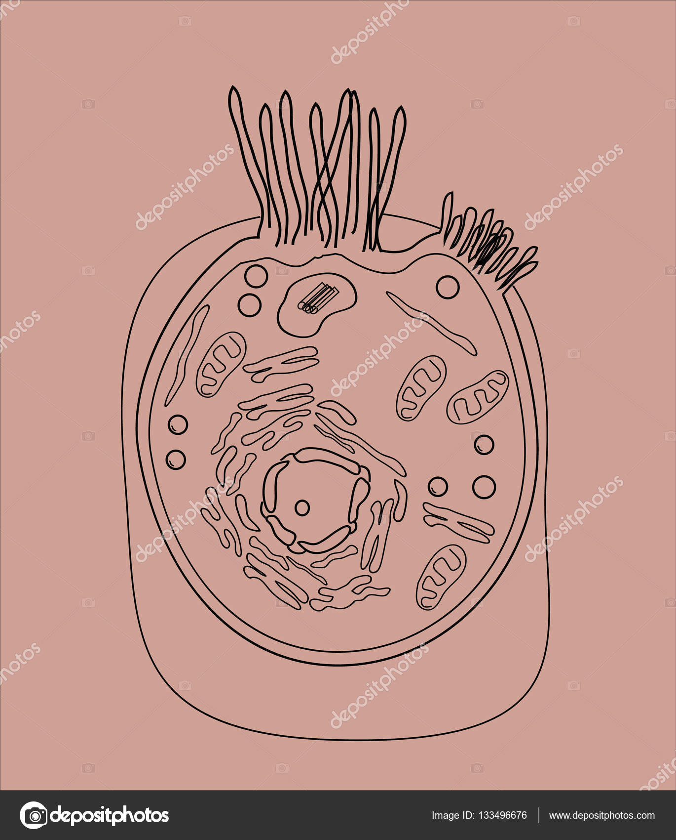
Human Cell Draw, Label, Define.docx - Human Cell Draw,... Human Cell Draw, Label, and Define Golgi Apparatus - A cell organelle found in the cytoplasm that synthesizes carbohydrates and packages materials for secretion from the cell. Chromatin - A thread like structure of genetic material from a cell that is not dividing Nucleus-Membrane part of cell that contains the hereditary material in chromosomes. 311,515 Human Cell Images, Stock Photos & Vectors | Shutterstock Find Human cell stock images in HD and millions of other royalty-free stock photos, illustrations and vectors in the Shutterstock collection. Thousands of new, high-quality pictures added every day. Human Heart Diagram Labeled | Science Trends The human heart is an organ responsible for pumping blood through the body, moving the blood (which carries valuable oxygen) to all the tissues in the body. Without the heart, the tissues couldn't get the oxygen they need and would die. Along with lymphatic vessels, the blood, blood vessels, and lymph, the heart composes the circulatory ... human cell diagram to label cell label shutterstock animal human structure labels organ vectors amp. Questions And Answers On Labeled/Unlebled Diagrams Of A Human Cell danauranauu.blogspot.com. unlabeled labeled diagrams jb004 k12 imageservice goconqr stilinterpretationen ausprobieren einrichtungsideen btec repetition vacuole iliaswasnua10.
Simple Columnar Epithelium: A Labeled Diagram and Functions These form a brush border. They also increase the absorptive surface area of these cells. On a concluding note, simple columnar epithelium has two primary functions of absorption and secretion. In the small intestine, it facilitates the absorption of nutrients. It also secretes mucus, which helps to lubricate, moisten, and protect the surface. The Human Skeleton: All You Need to Know - Bodytomy Bones are mostly confused as dead cells, but the human body skeleton consists of living cells, blood vessels and nerves. The point where two bones meet results in a joint that helps in the movement of the body. The muscles and ligaments are attached to the bones. The contraction and relaxation of the muscles and ligaments along with the joints ... Anatomy and Physiology: Parts of a Human Cell - Visible Body Cells can be divided into four groups: somatic, gamete, germ, and stem. Somatic cells are all the cells in the body that aren't sex cells, like blood cells, neurons, and osteocytes. Gametes are sex cells that join together during sexual reproduction. Germ cells produce gametes. Human Cells for Kids - Worksheet, Cell Model Activity, Review Game Human Body Printables makes a printable book that teaches students all about their heart, brain, muscles, cells, skin, bones, lungs, stomach, intestines, and bladder 4 EPIC Skeletal System Project ideas for kids - using things like pasta, life saver gummies, lego, and more you can learn about the human body in a fun memorable way
Life Science- Animal Cell- Labeling Diagram | Quizlet
Types of cells in the human body: Histology | Kenhub The most important types of cells are listed below. This article will discuss the histology of most important types of cells in the human organism. Contents Stem cells Red blood cells White blood cells Neutrophils Eosinophils Basophils Lymphocytes Platelets Nerve cells Neuroglial cells Muscle cells Skeletal muscle cells Cardiac muscle cells

Human Cell, Anatomy Image. 2 D Illustration, On White ...
human cell diagram with labels Human Body Parts | Anatomy System - Human Body Anatomy Diagram And anatomysystem.com. depicts. Onion Epidermal Cell Labeled - Top Label Maker labels-top.com. cell labeled onion epidermal elodea leaf cells labels lab function diagram microscope label salt every manual following slide visible living. Lower Leg Bones 1024×1350 | Anatomy System ...
Cytoplasm - Wikipedia
Human Cell Label Worksheet Teaching Resources | Teachers Pay Teachers Especially great for a science teacher's door decoration.Learning about the skeletal system and the human body? This is terrific for learning the bones of the human body skeleton. Print this 40 inch tall skeleton on any color 8.5x11in paper. Students easily cut and assemble the skeletal system with labels using the numbered tabs.

human cell Flashcards | Quizlet
Skeletal System - Labeled Diagrams of the Human Skeleton - Innerbody Each bone is a complex living organ that is made up of many cells, protein fibers, and minerals. The skeleton acts as a scaffold by providing support and protection for the soft tissues that make up the rest of the body. The skeletal system also provides attachment points for muscles to allow movements at the joints.
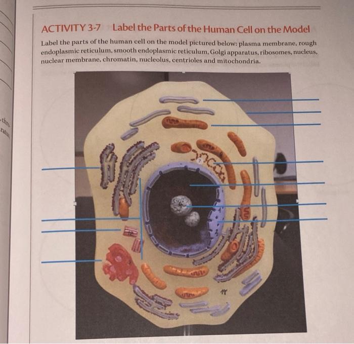
Solved ACTIVITY 3-7 Label the Parts of the Human Cell on the ...
Human Physiology - Cell structure and function Cell, or Plasma, membrane - encloses every human cell Structure - 2 primary building blocks include protein (about 60% of the membrane) and lipid, or fat (about 40% of the membrane). The primary lipid is called phospholipid , and molecules of phospholipid form a 'phospholipid bilayer' (two layers of phospholipid molecules).
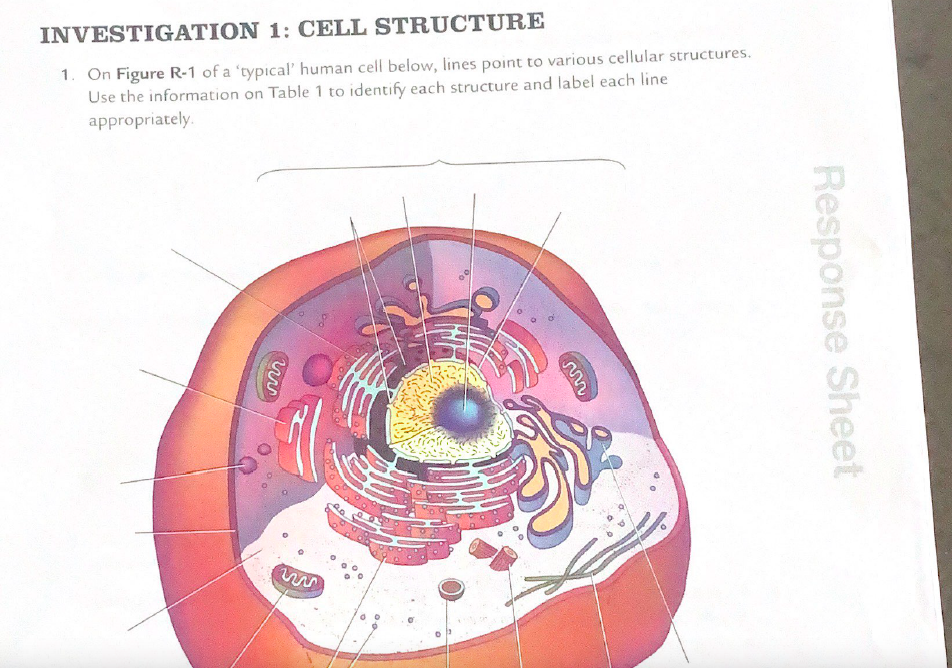
Solved INVESTIGATION 1: CELL STRUCTURE 1. On Figure R-1 of a ...
generalized human cell, labeled Diagram | Quizlet Start studying generalized human cell, labeled. Learn vocabulary, terms, and more with flashcards, games, and other study tools. Scheduled maintenance: Saturday, September 10 from 11PM to 12AM PDT

What Is Going On Inside That Cell? | Human cell diagram, Cell ...
Label Human Cell Teaching Resources | Teachers Pay Teachers Human Cells and Blood Cells ; Label the Various Cells Diagram:This is a great supplement for students to review/assess and strengthen their knowledge the on the TYPES HUMAN CELLS UNIT. Answer key is included. Colorful and Black and White versions of worksheet is included.It includes total ONE worksheet.

19,291 Human Cell Vector Illustrations & Clip Art - iStock
Anatomy and Physiology: Parts of a Human Cell

What is a cell? – YourGenome
Solved Select all of the following that are tenets of the ...
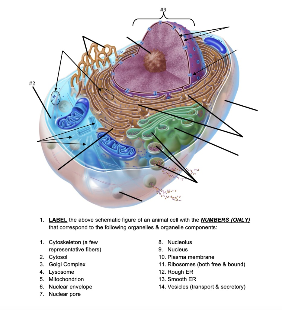
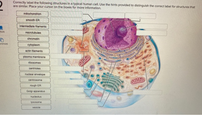
SOLVED: LABEL the above schematic figure of an animal cell ...

Label the Animal Cell Worksheets (SB11866) | Animal cells ...
Human Cell
Anatomy and Physiology: Parts of a Human Cell

Labelled Diagram Of A Human Cell Bone Cell Labeled Diagram ...
The structure of the human cells in a graphic image, cell ...
Anatomy and Physiology: Parts of a Human Cell
Cell Structure and Function Part 1 – The Organelles - Medical ...
Life Science - Lessons - Blendspace
Anatomy and Physiology: Parts of a Human Cell

Allen Integrated Cell Released Online

Human Cell Organelles Labeling Diagram | Quizlet

Animal cell anatomy. vector diagram. The structure of a ...
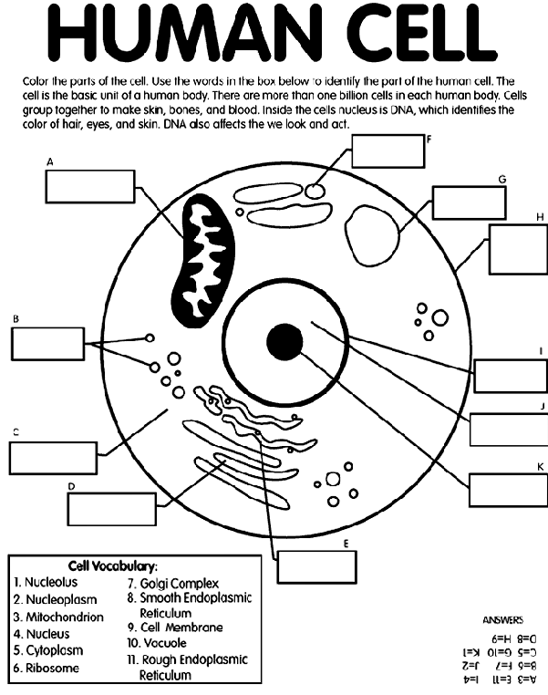
Human Cell Coloring Page | crayola.com
Label the human cell | Teaching Resources
Human Cell Diagram, Parts, Pictures, Structure and Functions ...

3D Rendering of the Human Cell Cross Section Stock ...
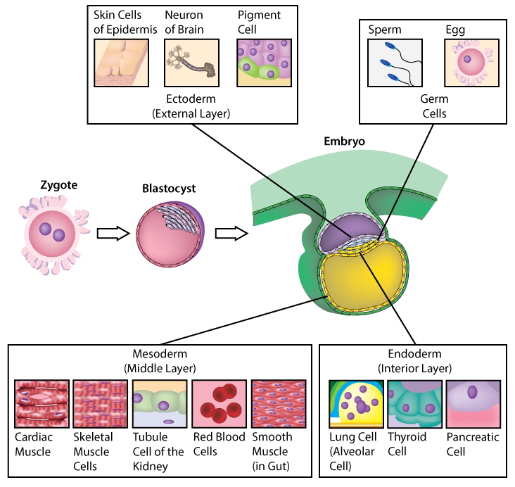
Human Biology fig. 1.107 - Cell Layers of the Embryo ...

Label the indicated structures in this diagram for eukaryotic ...
8,726 Cytoplasmic Images, Stock Photos & Vectors | Shutterstock

Human Cell Felt Board Human Cell Felt Set Human Cell - Etsy ...

4,485 Human Cell Diagram Stock Photos, Pictures & Royalty ...
cell | Definition, Types, Functions, Diagram, Division ...

Cells: The Building Blocks of Life | Human cell diagram, Cell ...

Cell Labeling - an overview | ScienceDirect Topics
Human cell anatomy realistic infographics with labelled ...

cell structure | Cell diagram, Human cell diagram, Animal ...
Post a Comment for "38 human cell with labels"