38 plastids diagram with labels
Plant Cells: Labelled Diagram, Definitions, and Structure - Research Tweet The cell wall is made of cellulose and lignin, which are strong and tough compounds. Plant Cells Labelled Plastids and Chloroplasts Plants make their own food through photosynthesis. Plant cells have plastids, which animal cells don't. Plastids are organelles used to make and store needed compounds. Chloroplasts are the most important of plastids. Plastids: Definition, Structure, Types, Functions and Diagram All kinds of green plastids are found to be surrounded by two membranes, these are inner and outer and are 7 nm thick membranes. They are separated by an 8-10nm thick periplastid space. A fully developed plastid's inner membranes do not have any inward foldings, unlike Mitochondria. Structure of Plastids Inheritance of Plastids
Plant Cell Diagram And Label : Draw A Diagram Of A Plant Cell And Label ... Draw A Diagram Of A Plant Cell And Label It S Any Four Parts Studyrankersonline from I'll also label the diagram. We think this is the most useful anatomy picture. We think this is the most useful anatomy picture.

Plastids diagram with labels
Chloroplast Structure and Function in detail with Labelled Diagram The chloroplasts are the cell organelles which consist of these pigments. The 3 types of pigments present in plants are chlorophyll, carotenoids, and anthocyanins. Chlorophyll imparts the green color to plants. Plastids are membrane-bound cytoplasmic organelles that can be found in the cells of plants and algae. Localization and distribution of YFP-labeled plastids in elongating ... Download scientific diagram | Localization and distribution of YFP-labeled plastids in elongating pollen tubes. (A) Plastids in pollen tubes of ACT1p::TP FtsZ1-YFP plants. YFP fluorescence or ... homeschoolgiveaways.com › 2022 › 06Free Printable Plant and Animal Cells Worksheets Jun 27, 2022 · Animal Cell Diagram. Worksheets of animal cell diagrams help your students to visually see what the animal cell looks like and identify visually the parts that make up the animal cell. Blank, Labeled, and Coloring Animal Cell Diagram – Grab these three free diagrams. One is labeled for studying and reference, the second is labeled but needs ...
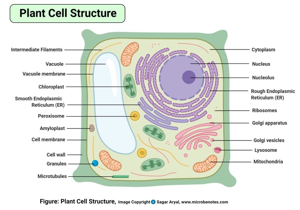
Plastids diagram with labels. Plant Cell-Definition, Structure, Parts, Functions, Labeled Diagram Figure: Labeled diagram of a plant cell, created with biorender.com The plant cell is comprised of cellulose, hemicellulose, and pectin, as well as plastids, which are essential for photosynthesis and starch storage, and enormous vacuoles that control cell turgor pressure. Labeled Plant Cell With Diagrams | Science Trends The parts of a plant cell include the cell wall, the cell membrane, the cytoskeleton or cytoplasm, the nucleus, the Golgi body, the mitochondria, the peroxisome's, the vacuoles, ribosomes, and the endoplasmic reticulum. Parts Of A Plant Cell The Cell Wall Let's start from the outside and work our way inwards. Plastids- Definition, Structure, Types, Functions and Diagram Plastid is a double membrane-bound organelle involved in the synthesis and storage of food, commonly found within the cells of photosynthetic plants. Plastids were discovered and named by Ernst Haeckel, but A. F. W. Schimper was the first to provide a clear definition. en.wikipedia.org › wiki › ChloroplastChloroplast - Wikipedia A chloroplast / ˈ k l ɔːr ə ˌ p l æ s t,-p l ɑː s t / is a type of membrane-bound organelle known as a plastid that conducts photosynthesis mostly in plant and algal cells.The photosynthetic pigment chlorophyll captures the energy from sunlight, converts it, and stores it in the energy-storage molecules ATP and NADPH while freeing oxygen from water in the cells.
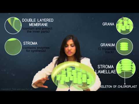
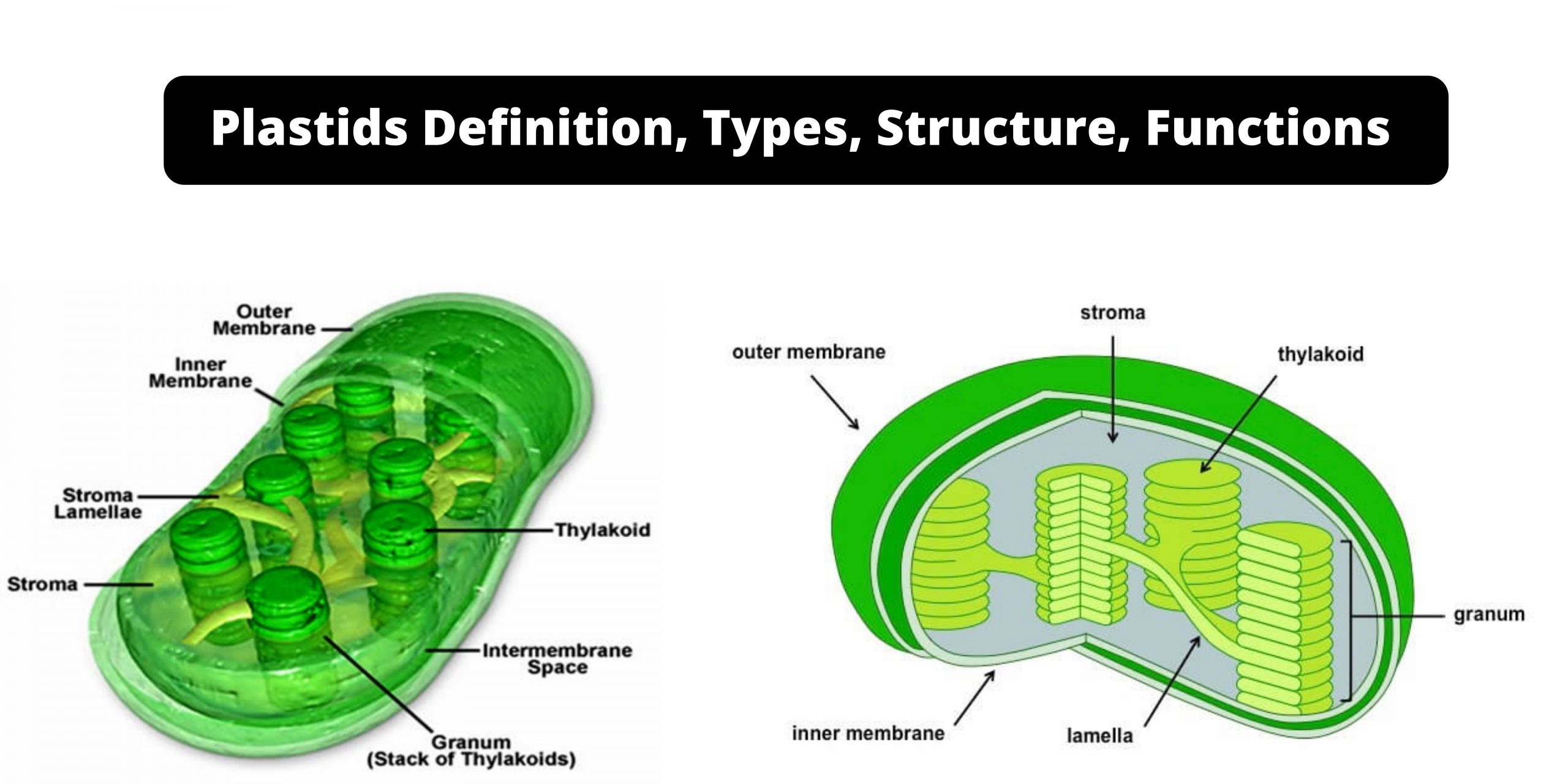
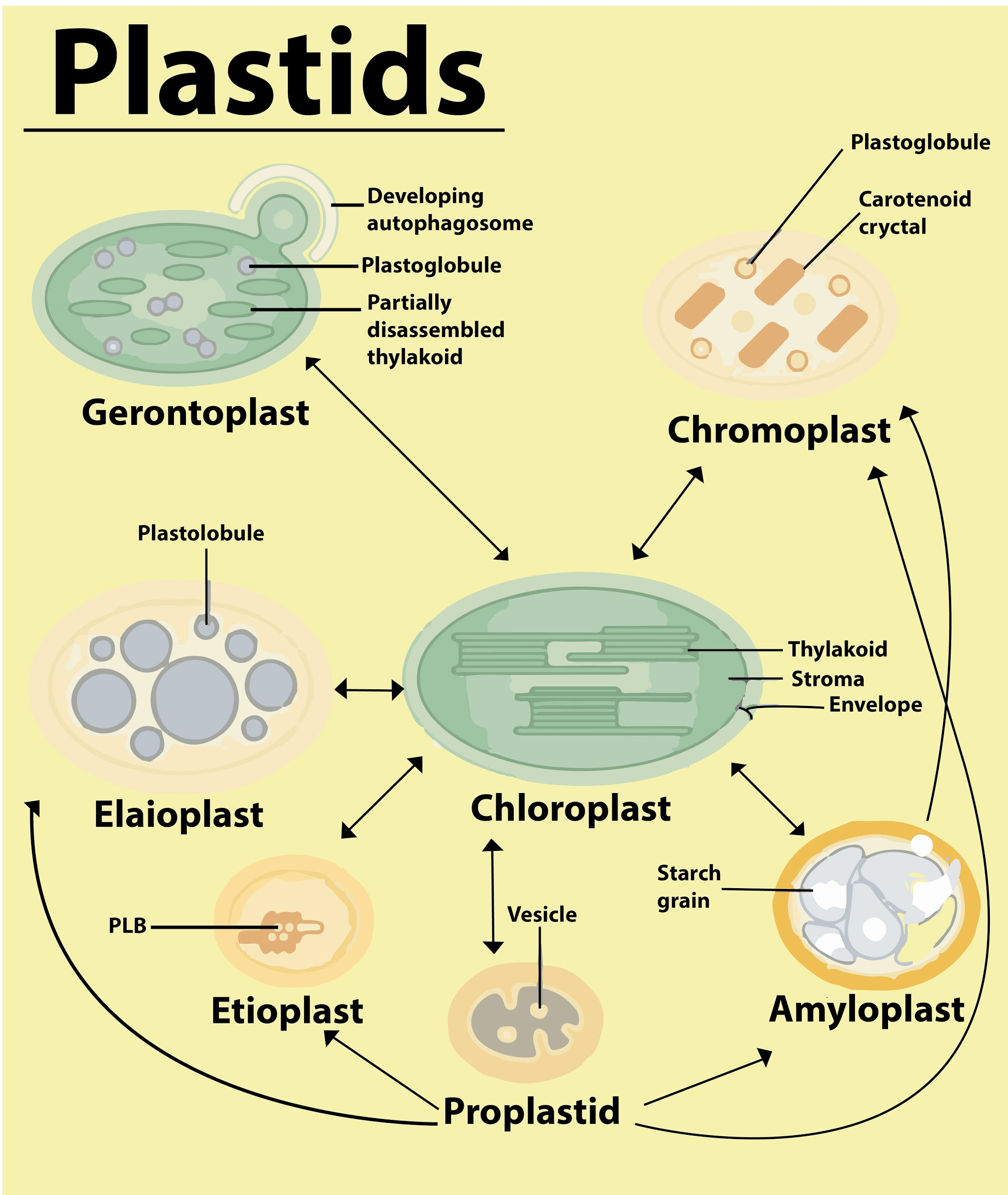
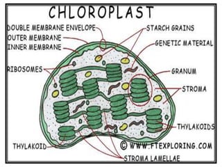
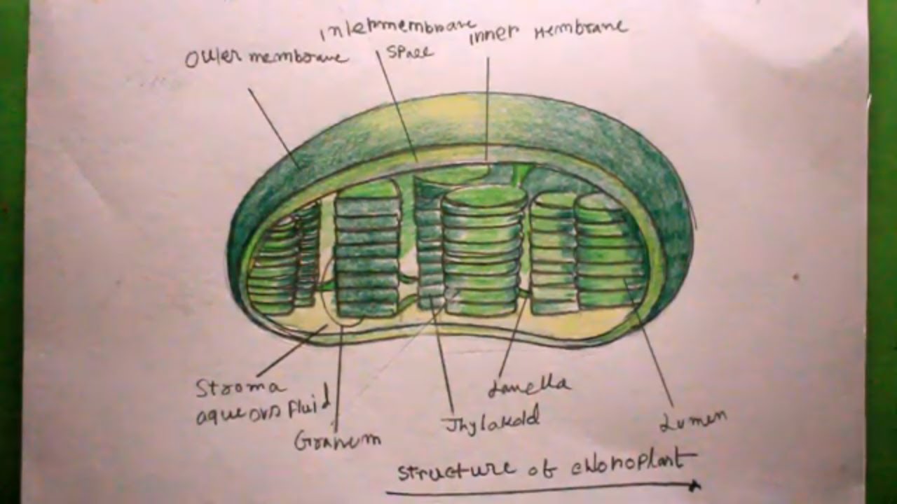
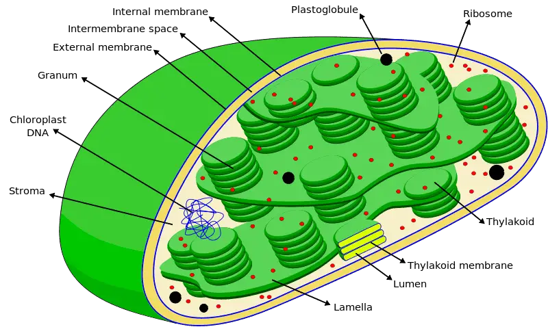
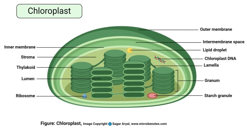
Plastids: Definition, Diagram, Types, and Plastid Function - Embibe Types of Plastids and Functions of Plastids Plastids are further divided into 3 types that have different functions and some have biological pigments as well. 1. Leucoplasts 2. Chromoplasts 3. Chloroplasts Leucoplasts These colourless Plastids possess internal lamellae and do not contain photosynthetic and grana pigments. Chloroplast- Diagram, Structure and Function Of Chloroplast - BYJUS The chloroplast diagram below represents the chloroplast structure mentioning the different parts of the chloroplast. The parts of a chloroplast such as the inner membrane, outer membrane, intermembrane space, thylakoid membrane, stroma and lamella can be clearly marked out. Chloroplast Diagram representing Chloroplast Structure Plastids: Types, Structure and Function (With Diagram) - Biology Discussion Plastids may be coloured or colourless and are of three types. The leucoplasts are the colourless plastids principally serving the purpose of storage. On the basis of nature of storage compound, leucoplastids are amyloplasts (starch), elaioplasts (oil) or aleuroplasts (protein). The green plastids or chloroplastids are needed for photosynthesis. agl.freinet.es › click-and-drag-each-label-toClick And Drag Each Label To Identify The Organelles ... Drag the correct labels onto the diagram to identify the structures and molecules involved in translation Solved carbon dioxide transport drag each label to the ap Solved carbon dioxide transport drag each label to the ap. Apr 13, 2017 · Drag the labels onto the diagram to identify the stages of cellular respiration Drag the labels on the left.
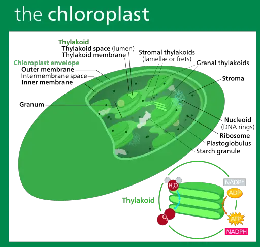
› pmc › articlesStructure of 3-oxoacyl-(acyl-carrier protein) synthase II ... May 01, 2008 · The amino-acid sequences of KAS I and KAS II share 38% identity and their functions are closely related, catalyzing the condensation of acyl-ACP with malonyl-ACP. In plastids, KAS I extends C 4 in the substrate to C 16 in six rounds of elongation, whereas KAS II carries out an additional step to give a C 18 product (Olsen et al., 2001 ). History of botany - Wikipedia Botany (Greek Βοτάνη - grass, fodder; Medieval Latin botanicus – herb, plant) and zoology are, historically, the core disciplines of biology whose history is closely associated with the natural sciences chemistry, physics and geology.A distinction can be made between botanical science in a pure sense, as the study of plants themselves, and botany as applied science, which studies … Plant and Animal Cell: Labeled Diagram, Structure, Function - Embibe Plastids: 1. Double membrane-bound structures found only in the plant cells. 2. This is an autonomous organelle. 3. There are stroma or matrix and grana or stacked discs that are involved in photosynthesis. 4. Grana are the site for photochemical reactions of photosynthesis, while stroma is the site for biochemical reactions of photosynthesis. 5. Cell Organelles- Definition, Structure, Functions, Diagram However, some organelles are specific to one particular type of cell-like plastids and cell walls in plant cells. Image created using biorender.com. List of Cell Organelles. Cell membrane (Plasma membrane/ Plasmalemma) ... Labeled Diagram; Prokaryotes vs Eukaryotes- Definition, 47 Differences, Structure, Examples; Amazing 27 Things Under The ...
File:Plastids types en.svg - Wikimedia Commons
› lecture › algaeAlgae as a Photosynthetic Organism - Algae Basics | Coursera Sep 04, 2019 · So, the diagram on the right represents the chloroplast, but also in many ways would represent the fundamental structure, and function of a cyanobacterium. Digging down even at a finer scale, photosynthesis at the molecular scale. On the left is a diagram of the membrane, which is the linear pattern that you see there.

draw a well labelled diagram of a plastid? where is it found ...
› books › NBK3841Gene Help: Integrated Access to Genes of Genomes in the ... Sep 13, 2006 · The gene being shown on the diagram is in maroon. All other diagrams and labels anchor links to specific Gene pages, supporting quick navigation to review neighboring genes by clicking in the area of the symbol/arrow. The diagram shows the gene’s placement on any and all chromosomes in the current genome annotation.
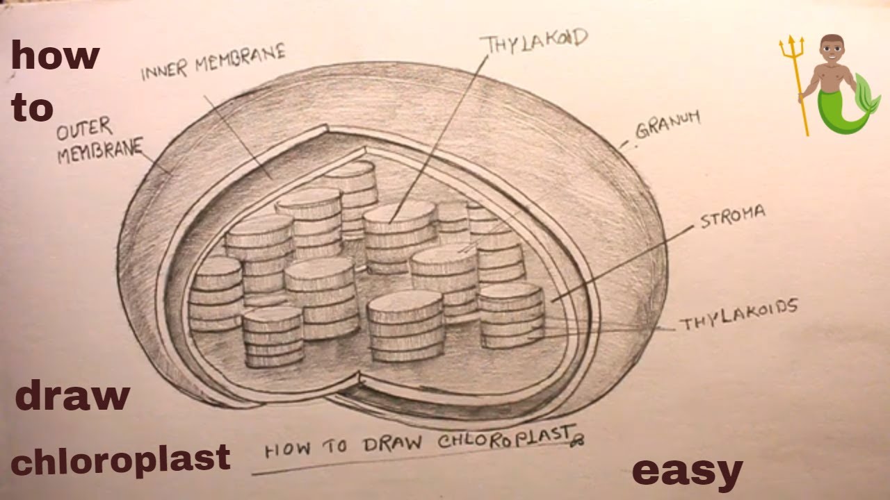
How TO Draw plastid/draw chloroplast/chloroplast drawing
Plastids - Different types of Plastids and their functions in ... - BYJUS There are different types of plastids with their specialized functions. Among them, a few are mainly classified based on the presence or absence of the Biological pigments and their stages of development. Chloroplasts Chromoplasts Gerontoplasts Leucoplasts Chloroplasts

Differentiation of chromoplasts and other plastids in plants ...
dokumen.pub › campbell-biology-12th-edition-12Campbell Biology, 12th Edition [12nbsped.] 9780135988046 ... In Chapter 12, the cell cycle figure (Figure 12.6) now includes cell images and labels describing the events of each phase. Unit 3 GENETICS Chapters 13–17 incorporate changes that help students grasp the more abstract concepts of genetics and their chromosomal and molecular underpinnings.
With a neat labelled diagram explain the ultra structure of ...
Stanford University UNK the , . of and in " a to was is ) ( for as on by he with 's that at from his it an were are which this also be has or : had first one their its new after but who not they have

Which type of plastids help in photosynthesis? Draw its diagram.
Plastids: Everything You Need to Know and More Plastids are a diverse group of double-membrane bound organelles found in most plants and algae. They may also be found in ferns, moss, parasitic worms and marine mollusks. Apart from photosynthesis, these organelles also assist in food storage and synthesis of compounds such as lipids, amino acids and carbohydrates.

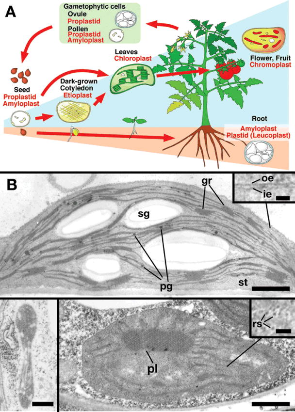
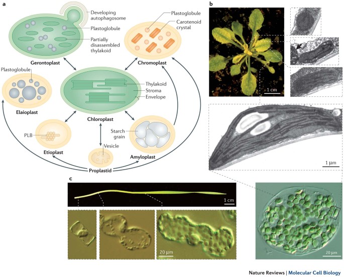
Diversity of plastid types and their interconversions ...
The Stanford Natural Language Processing Group ' '' ''' - -- --- ---- ----- ----- ----- ----- ----- ----- ----- ----- ----- ----- ----- ----- ----- ----- ----- ----- ----- ----- ----- ----- ----- ----- ----- ----- ----- ----- ----- ----- ----- ----- ----- ----- ----- ----- ----- ----- ----- ----- ----- ----- ----- ----- ----- ----- ----- ----- ----- ----- ----- ----- ----- ----- ----- ----- ----- ----- -----
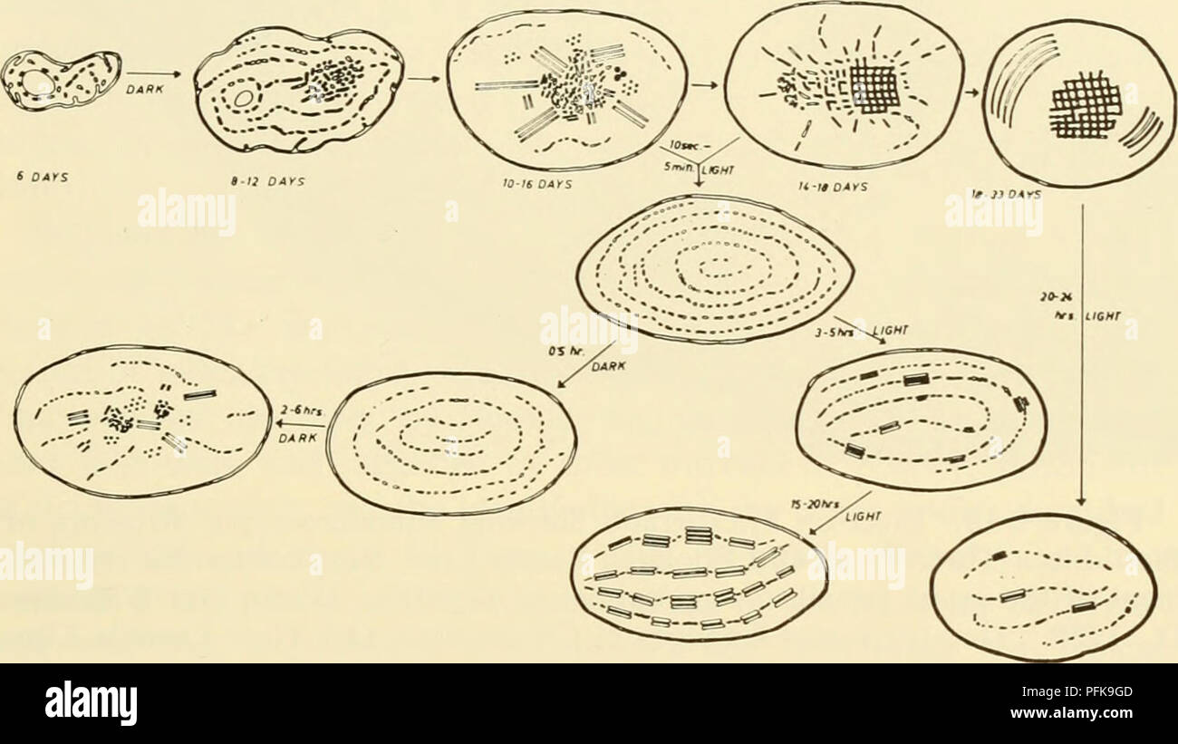
Cytology. Cytology. plastids increase in size but do not ...
Study Notes on Genetics of Plastids (With Diagram) - Biology Discussion The below mentioned article provides a study note on Genetics of Plastids. In the cytoplasm of the plant cells are found many small cytoplasmic bodies, called plastids. These plastids are of several types, such as, chloroplast, leucoplast, chromoplast and so on. Plastids arise from smaller particles, the proplastids, found in the egg cytoplasm.

Chloroplast - Wikipedia
EOF
Plant Cell- Definition, Structure, Parts, Functions, Labeled ...
Plastid - Wikipedia There is an illustration of stages depicted by the diagram mentioned above in which it is shown inter-conversion of Plastids In 1977 J.M Whatley proposed a plastid development cycle which said that plastid development is not always unidirectional but is a cyclic process several times.

Plastids and it's types/ Chloroplast in detail
homeschoolgiveaways.com › 2022 › 06Free Printable Plant and Animal Cells Worksheets Jun 27, 2022 · Animal Cell Diagram. Worksheets of animal cell diagrams help your students to visually see what the animal cell looks like and identify visually the parts that make up the animal cell. Blank, Labeled, and Coloring Animal Cell Diagram – Grab these three free diagrams. One is labeled for studying and reference, the second is labeled but needs ...

3D Diagram Of A Plant Cell | World of Reference | Plant cell ...
Localization and distribution of YFP-labeled plastids in elongating ... Download scientific diagram | Localization and distribution of YFP-labeled plastids in elongating pollen tubes. (A) Plastids in pollen tubes of ACT1p::TP FtsZ1-YFP plants. YFP fluorescence or ...

Plastids in Plant Cells Function & Types | What do Plastids ...
Chloroplast Structure and Function in detail with Labelled Diagram The chloroplasts are the cell organelles which consist of these pigments. The 3 types of pigments present in plants are chlorophyll, carotenoids, and anthocyanins. Chlorophyll imparts the green color to plants. Plastids are membrane-bound cytoplasmic organelles that can be found in the cells of plants and algae.

how to draw chloroplast/draw chloroplast diagram easily
Chloroplast Biogenesis: Control of Plastid Development ...

Plastids
Biogenesis and homeostasis of chloroplasts and other plastids ...

Evolution of plastid involving peptidoglycan. Plastids arose ...
Plastids - Different types of Plastids and their functions in ...
Plastids Definition, Types, Structure, Functions

Plastid Diagram | Types |Structure | Function | Structure and ...
Plastids - Definition, Types, Main Structure and Function
Plastids are found in aAll animal cells bSome animals class ...

Plastids - Introduction, Types and Functions

Transport of Proteins into Cryptomonads Complex Plastids ...

Plastids
Plastids

Plastid and its various types with their respective organelle ...
How TO Draw plastid/draw cloroplast easy/cloroplast drawing for science project

Plastids in Plant Cells Function & Types | What do Plastids ...

Endosymbiotic origin of the Archaeplastida plastid through ...

draw a well labeled diagram of a plastid where it is found ...
Plastid Black and White Stock Photos & Images - Alamy

The Chlamydomonas plastid testbed. The design stage of the ...

What are the Functions of Plastids – Techy Bois
Plastids: Everything You Need to Know and More – Microscope ...
Plant Cell- Definition, Structure, Parts, Functions, Labeled ...

Plastids in a cell - function, types, structure and meaning
Post a Comment for "38 plastids diagram with labels"