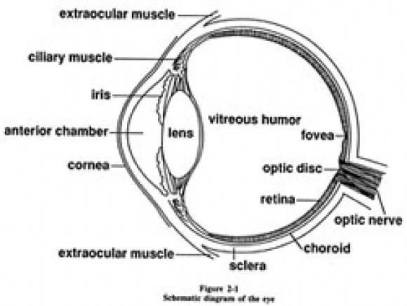
39 diagram of the human eye without labels
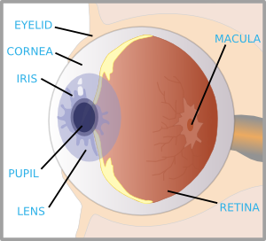
Label the Eye Diagram - Enchanted Learning Label the Eye Diagram. Human Anatomy. Read the definitions, then label the eye anatomy diagram below. Cornea - the clear, dome-shaped tissue covering the front of the eye. Iris - the colored part of the eye - it controls the amount of light that enters the eye by changing the size of the pupil. Lens - a crystalline structure located just behind ... Human Body Parts Images Without Labels - Free Vector Download 2020 Find human body part labels stock images in hd and millions of other royalty free stock photos illustrations and vectors in the shutterstock collection. The vagina and vulva are important but often misunderstood parts of the human body. Posted in bones diagrams tagged body skeleton. Human body parts pictures with names.
Eyes - Layers of Learning | Human eye diagram, Parts of ... Elementary Science. Description Use these simple eye diagrams to help students learn about the human eye. Three differentiated worksheets are included: 1. Write the words using a word bank 2. Cut and paste the words 3. Write the words without a word bank Labels include: eyebrow, eyelid, eyelashes, pupil, iris, and sclera.
Diagram of the human eye without labels
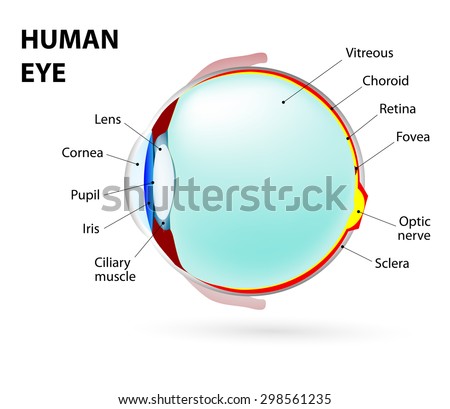
PDF Parts of the Eye - National Eye Institute | National Eye Institute Eye Diagram Handout Author: National Eye Health Education Program of the National Eye Institute, National Institutes of Health Subject: Handout illustrating parts of the eye Keywords: parts of the eye, eye diagram, vitreous gel, iris, cornea, pupil, lens, optic nerve, macula, retina Created Date: 12/16/2011 12:39:09 PM Anatomy of the eye: Quizzes and diagrams - Kenhub Take a look at the diagram of the eyeball above. Here you can see all of the main structures in this area. Spend some time reviewing the name and location of each one, then try to label the eye yourself - without peeking! - using the eye diagram (blank) below. Unlabeled diagram of the eye Eye Anatomy: 16 Parts of the Eye & Their Functions The lens of the eye (or crystalline lens) is the transparent lentil-shaped structure inside your eye. This is the natural lens. It is located behind the iris and to the front of the vitreous humor (vitreous body). The vitreous humor is a clear, colorless, gelatinous mass that fills the gap between the lens and the retina in the eye.
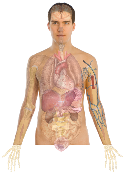
Diagram of the human eye without labels. PDF Eye Anatomy Handout - National Eye Institute of light entering the eye. Lens: The lens is a clear part of the eye behind the iris that helps to focus light, or an image, on the retina. Macula: The macula is the small, sensitive area of the retina that gives central vision. It is located in the center of the retina. Optic nerve: The optic nerve is the largest sensory nerve of the eye. Human Body Diagram - Bodytomy ☛ The human eye has the ability to differentiate between 400+ shades of gray, and what's more, it can identify approximately 10 million colors. ☛ Your ears never sleep. Sound is received even while you are asleep; it's the brain that does not process them. Human Ear Diagram - Bodytomy The Structure of Human Ear. Helix: It is the prominent outer rim of the external ear. Antihelix: It is the cartilage curve that is situated parallel to the helix. Crus of the Helix: It is the landmark of the outer ear, situated right above the pointy protrusion known as the tragus. Auditory Ossicles: The three small bones in the middle ear ... Cross sectional diagram of human eye [1]. | Download ... Context in source publication. ... eye is housed in a socket of bone called the orbit and is pro- tected from the external air by the eyelids [9]. A cross section of the eye is shown in Figure 1 ...
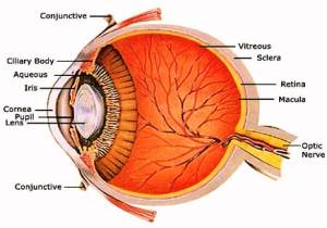
The Eyes (Human Anatomy): Diagram, Optic Nerve, Iris ... Articles On Eye Basics. Your eye is a slightly asymmetrical globe, about an inch in diameter. The front part (what you see in the mirror) includes: Iris: the colored part. Cornea: a clear dome ... Eye Diagram With Labels and detailed description - BYJUS A brief description of the eye along with a well-labelled diagram is given below for reference. Well-Labelled Diagram of Eye The anterior chamber of the eye is the space between the cornea and the iris and is filled with a lubricating fluid, aqueous humour. The vascular layer of the eye, known as the choroid contains the connective tissue. Structure and Functions of Human Eye with labelled Diagram The human eye is a roughly spherical organ, responsible for perceiving visual stimuli. It is enclosed within the eye sockets in the skull and is anchored down by muscles within the sockets. Anatomically, the eye comprises two components fused into one; hence, it does not possess a perfect spherical shape. Eye Diagram Unlabelled - Wiring Diagram Pictures Best Human eye diagram unlabelled free vector download for commercial use in ai, eps, cdr, svg vector illustration graphic art design format. human eye. Ask A Biologistcoloring page | Web address:schematron.org coloring. Human Eye. Page 2. 5. 3. 2. 4. How to draw human eye in easy steps -10th -Physics - science - CBSE syllabus - NCERT class 10
Eye Diagram Teaching Resources | Teachers Pay Teachers Anatomy of the Eye Diagrams for Coloring/Labeling, with Reference and Summary by Homemade For Play 7 $1.95 PDF This printable contains 13 clear and simple cross sectional diagrams of the human eye. Eye Diagram - Differentiated Worksheets and ... - Pinterest Eye Diagram - Differentiated Worksheets and EASEL Activities Description Use these simple eye diagrams to help students learn about the human eye. Three differentiated worksheets are included: 1. Write the words using a word bank 2. Cut and paste the words 3. Eye Anatomy: 16 Parts of the Eye & Their Functions The lens of the eye (or crystalline lens) is the transparent lentil-shaped structure inside your eye. This is the natural lens. It is located behind the iris and to the front of the vitreous humor (vitreous body). The vitreous humor is a clear, colorless, gelatinous mass that fills the gap between the lens and the retina in the eye. Anatomy of the eye: Quizzes and diagrams - Kenhub Take a look at the diagram of the eyeball above. Here you can see all of the main structures in this area. Spend some time reviewing the name and location of each one, then try to label the eye yourself - without peeking! - using the eye diagram (blank) below. Unlabeled diagram of the eye
Anatomy And Physiology Of The Eye - Anatomy Drawing Diagram
PDF Parts of the Eye - National Eye Institute | National Eye Institute Eye Diagram Handout Author: National Eye Health Education Program of the National Eye Institute, National Institutes of Health Subject: Handout illustrating parts of the eye Keywords: parts of the eye, eye diagram, vitreous gel, iris, cornea, pupil, lens, optic nerve, macula, retina Created Date: 12/16/2011 12:39:09 PM

OMTEX CLASSES: Science and Technology Diagram for SSC Maharashtra State Board

Human Eye Model - YouTube
Eye Diagram - Cliparts.co

Human Eye Diagram Labeled - Health, Medicine and Anatomy Reference Pictures | A&P | Pinterest ...
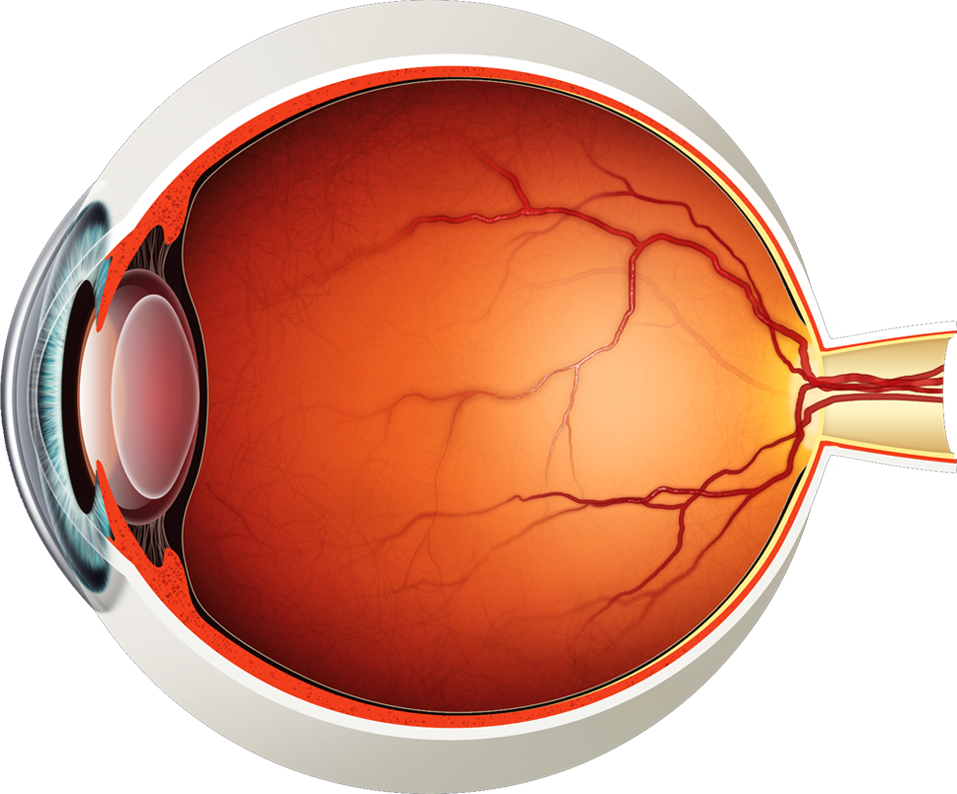
Diagram of the Eye - Lions Eye Institute
eye anatomy | Anatomy System - Human Body Anatomy diagram and chart images

Draw And Label A Human Eye

Towson Physics with Sean Lally: Optics - Camera and Eye

Eye diagram
Schematic Diagram Eye Human Anatomy Labeled Stock Illustration 298561235 - Shutterstock
Human Body Anatomy Basics No Lines Clip Art at Clker.com - vector clip art online, royalty free ...

Schematic Diagram Of The Eye. Human Anatomy. Labeled Stock Photo 298561235 : Shutterstock

Structure of the human eye. | Download Scientific Diagram

Medical Stock Art, Anatomy of the Eye
Eye With Labels Clip Art at Clker.com - vector clip art online, royalty free & public domain

picture front of the eye without labels clipart - Clipground

Daniela's Biology Lab
Post a Comment for "39 diagram of the human eye without labels"